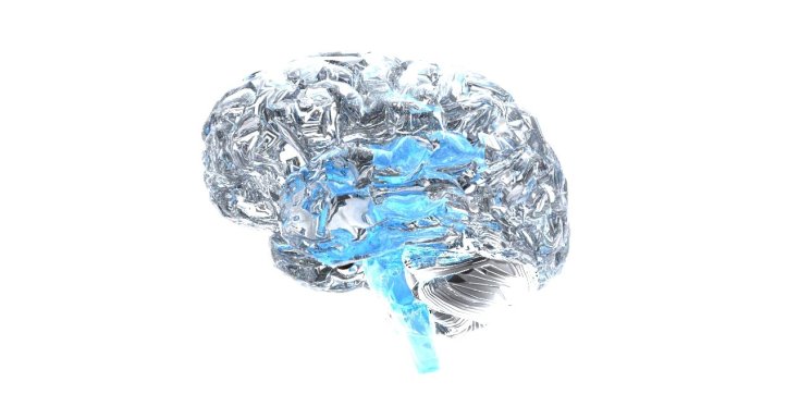
The paper reviewed here is ‘Roles of the Insular Cortex in the Modulation of Pain: Insights from Brain Lesions’ by Starr and colleagues and freely available here. This is of relevance to a model of the insular cortex and emotions which i’ve been working on (rather slowly!). The researchers have identified two subjects who had developed middle cerebral artery strokes with resulting right sided insular lesions. They compare these subjects with a healthy control group and examine how pain is processed using a number of different methodologies.
Methodology
The methodology is complex as the study consists of multiple components – assessments of pain intensity, the unpleasantness of the pain, a characterisation of the individual responses to the pain and a between group comparison. 2 subjects with strokes involving the left Insular cortex were identified. They were compared with 14 healthy controls with a mean age of 59. The subjects themselves were aged 53 and 59. If the control group were healthy then I interpreted this as meaning that there was no significant medical illness. However conditions such as hypertension and diabetes are more prevalent with increasing age and it could be argued therefore that since the controls are reported as ‘healthy’ then they may not be representative of the general population. Additionally there might be expected to be evidence of white matter lesions however small in at least some of the control group although these are not reported although if they did exist they might not be too important for the current paradigm.
Subjects were administered a visual analogue scale for pain intensity and pain unpleasantness. Short and long duration noxious stimuli were applied to the calf (as this was unaffected by the lesions in the two subjects). Tactile and thermal thresholds were also identified in all subjects. fMRI was used to investigate brain activation during presentation with stimuli. There were parts of the methodology I didn’t understand even after looking for further information. The difficulties I had were with the ‘statistical analysis of regional signal changes within the brain’. For ‘pain-related activations’, the researchers used ‘boxcar activations’. Boxcar functions are described here. This much is reasonably straightforward. The researchers are modelling the pain activations (presumably in the brain regions) as signals of finite duration with a specific and constant amplitude meaning that if graphed against time it looks like a rectangle with no activity on either side of the signal. The use of activations suggested to me that they were referring to brain activations, after all this part of the study is concerned with fMRI. They then refer to the regressor and here I wasn’t entirely clear which variable they were referring to. They add that
‘the regressor was convolved with a gamma-variate model for the hemodynamic response … and its temporal derivative‘
I hadn’t come across the term convolved before but it appears to mean that two functions are combined (there’s a good illustration here). I presume that the gamma-variate model is equivalent to a gamma-distribution which would be determined by the relevant parameters. The individual components make sense but I couldn’t put it all together. The regressor (what is the regressor in this case?) is combined with a model of the hemodynamic response. The hemodynamic response is the change in blood flow to the region of interest representing a proxy marker for cerebral activity but why is the regressor being combined with this response? Why for instance would they not be using a correlation between the VAS responses and the hemodynamic response (approximated with the gamma-variate model)? They further add that they are using a temporal derivative of the hemodynamic response. Presumably this would then refer to a change in change in blood flow (if the derivative is being used in the calculus sense) but why would they be interested in this? So, I couldn’t understand this section although the remainder of the paper was reasonably straightforward and in effect I had to then accept the results for this part of the paper without fully understanding the process. I have argued elsewhere that there is a place for having an accompanying video with papers (for instance that could be freely hosted on YouTube) and I suspect that a 5-minute video could save the reader at least 30 minutes of reading around the subject (or more) particularly when multiple methodologies (some esoteric) are being used. The end result is that when presented with the noxious stimuli, the researchers have used fMRI on the 2 subjects who have had a stroke and also the healthy control group to image where the activation is taking place. The methodology is more involved than that but I thought these were the most salient points.
Results
With a complex methodology there were lots of results. The ones I found most interesting were as follows
1. Patients 1 and 2 had involvement of the Sensory Cortex and Basal Ganglia as well as the Insular Cortex from the strokes.
2. Subjects 1 and 2 differed subtly in the regions that were affected
3. Subjects 1 and 2 had increased pain intensity (on the affected side) but lowered pain unpleasantness in response to the noxious stimuli in comparison to the control group
4. In the subjects, the affected insula was not activated in response to pain which the researchers reasonably suggest is due to the damage resulting from the stroke
5. In response to pain, in one of the subjects the prefrontal cortex is activated in contrast with the control group which the researchers suggest is being additionally recruited in processing pain without the insula being active.
6. Without going into the details too much, the researchers have identified somatosensory cortex activation on the side where the insula is not active which wasn’t present in the control group. They interpret this as meaning that this activity is not reliably picked up when the insular cortex is active. Does this mean that some areas mask others in fMRI studies?
Conclusions
On reading this paper, I thought that there were some obvious limitations which the authors acknowledge. Thus there are only 2 subjects and there are multiple areas involved – not just the insular cortex. However, the researchers were selecting for subjects that would have reasonably localised involvement of the insular cortex and the small sample size reflects the practical reality of recruitment for these very specific criteria. The involvement of multiple areas is characteristic of this paradigm and the evidence should be triangulated with evidence from elsewhere (which the researchers have done in the discussion). What I thought was slightly more tricky from the modelling perspective is that the subjects have separate responses both in terms of activation and also in terms of their responses to stimuli. However this could be a direct result of the above. In other words it they have slightly different patterns of injury then it is not unreasonable to suppose that they would have different consequences both in terms of conscious experience and therefore of the measurement of that experience. What is interesting from the modelling perspective is that some fairly broad conclusions can be drawn from this. There is some good evidence from this study that the insular cortex might be involved in gauging how unpleasant a pain stimulus is. Thus the subjects rated the pain as more intense on some occasions and yet they didn’t have a corresponding increase in the unpleasantness of that pain. This might also be described as a dissociation of the cognitive description of that pain and the emotional experience of that pain and elsewhere the insular cortex has been described as a region which is involved in the labelling of pain and emotional experiences. However that is not to say that the somatosensory cortex and basal ganglia might be involved in this process instead. The researchers though have arguments against these possibilities from their data. What is also interesting from a modelling perspective is the apparent collateral network that is invoked when the insular cortex doesn’t appear to be working properly. Thus the prefrontal cortex appears to be involved in one case suggesting that if one region is inactive there is possibly an automatic ‘redirect’ to an alternative region. Could the insular cortex be directly involved in tolerance of pain on the basis of these results? If so, then it might suggest that an understanding of the neurotransmitters used in this region might be of relevance to therapeutics in pain management. Additionally there may be implications for chronic illnesses where pain is a feature. Pain also has a complex relationship with depression. This part of the discussion is necessarily theoretical but this area thus has many possible ramifications.
Index
You can find an index of the site here. The page contains links to all of the articles in the blog in chronological order.
You can follow ‘The Amazing World of Psychiatry’ Twitter by clicking on this link
Podcast
You can listen to this post on Odiogo by clicking on this link (there may be a small delay between publishing of the blog article and the availability of the podcast).
TAWOP Channel
You can follow the TAWOP Channel on YouTube by clicking on this link
Responses
If you have any comments, you can leave them below or alternatively e-mail justinmarley17@yahoo.co.uk
Disclaimer
The comments made here represent the opinions of the author and do not represent the profession or any body/organisation. The comments made here are not meant as a source of medical advice and those seeking medical advice are advised to consult with their own doctor. The author is not responsible for the contents of any external sites that are linked to in this blog.

I can’t say I agree 100% on certain thoughts, but you certainly have a unique perspective. Anyway, I enjoy the quality you add to
LikeLike
[…] Role of the Insular Cortex in the Modulation of Pain […]
LikeLike
I visited various web pages however the audio feature for audio songs current at
this site is genuinely wonderful.
LikeLike