
Dr Korbinian Brodmann, German Neurologist, Frontpiece of ‘Localisation in the Cerebral Cortex’, 1909, Public Domain*
The Brain is a complex organ, responsible for the full gamete of our inner experiences whether these are our first thoughts on waking, the perception of a rainbow or the sharing of joy with others. Understanding the brain has been an almost unobtainable goal which many great scientists have striven for. One scientist who realised the immense complexity of the task set out to characterise the brain in a more limited way and in the process established one of the most successful maps of the brain which continues to be routinely used over 100 years later. His name was Dr Korbinian Brodmann. In the first part of this series, there was a brief look at the context of Brodmann’s landmark work ‘Brodmann’s Localisation in the Cerebral Cortex’. In the second part of the series we will take a closer look at the 21 pages of his book which relate to the special regions in the human Cerebral Cortex that Brodmann identified.
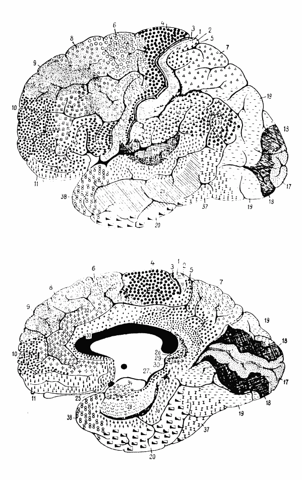
The central 21 pages of Brodmann’s work are contained within Chapter IV ‘Description of Individual Brain Maps’ in which he contrasts the brain maps of several species including humans. The third region he looks at is the Frontal region.

Cytoarchitectonics of human brain according to Brodmann (1909), Public Domain*
According to Brodmann, the Frontal Region is divided into eight subregions. Interestingly he contrasts this with the work of two other anatomists, Elliot Smith who identifies 8 subregions and Campbell who identies 2 subregions. Furthermore although Smith also identifies eight subregions, they are not the same 8 subregions that Brodmann identifies. For several subregions, Brodmann suggests that these can be further subdivided. For other subregions he suggests that they are very difficult to demarcate and that there is considerable individual variation.

Three drawings by Santiago Ramon y Cajal, taken from the book “Comparative study of the sensory areas of the human cortex”, pages 314, 361, and 363, Public Domain*
Left: Nissl-stained visual cortex Middle: Nissl-stained motor cortex Right: Golgi-stained cortex

Cingulate Sulcus, Finn Årup Nielsen, GNU Free Documentation License.
Area 8: Intermediate Frontal Area
Borders
Medial – Cingulate Sulcus
Lateral – Middle Frontal Gyrus
Area 9: The Granular Frontal Area
Borders
Medial – Cingulate Sulcus
Ventrolateral – Inferior Frontal Sulcus
Area 10: The Frontopolar Area
Borders
Inferiomedial – Superior Rostral Sulcus
Brodmann states that this area approximates the Frontal Area of Elliot Smith.
Area 11: The Prefrontal Area
Borders
Medial – Superior Rostral Sulcus
Lateral – Frontomarginal Sulcus of Wernicke
Orbital – Medial Orbital Sulcus
Area 44: The Opercular Area
Borders
Posterior – Inferior Precentral Sulcus
Superior – Inferior Frontal Sulcus
Anterior – Ascending Ramus of Sylvian Fissure
Inferiomedial – Frontal Operculum and Insular Cortex
Brodmann suggests that there is justification for an Anterior and Posterior Opercular Area.
Area 45: The Triangular Area
Borders
Caudal – Ascending Ramus of the Sylvian Fissure
Dorsal – Inferior Frontal Sulcus
Rostral – Radiate Sulcus of Eberstaller
Inferior – Insular Cortex
For areas 44 and 45, Brodmann states that there is considerable individual variation in the sulci.
Area 47: The Orbital Area
Situated along the Orbital Sulcus.
Border
Lateral – Crosses Orbital part of Inferior Frontal Gyrus
Brodmann suggested that this area could be combined with Area 44 and 45 to form a Subfrontal Subregion.
Area 46: The Middle Frontal Area
Brodmann states that there is an ambiguous demarcation because this area is not bounded by sulci.
The Middle Frontal Area extends to the anterior aspect of Inferior Frontal Gyrus as it reaches the Orbital surface and includes the middle third of the middle of the Inferior Frontal Gyrus
The Frontal Region is perhaps the most complex region described by Brodmann, being divided into eight subregions with complex and sometimes ambigous boundaries. Nevertheless this region plays a significant role in human personality and behaviour and Brodmann’s work has laid the neuroanatomical foundations for research into these functions.
References
Brodmann’s Localisation in the Cerebral Cortex. 1909. Translated and Edited by Laurence J Garey. Springer. 2006.
*Public Domain in those countries where the Copyright term of the life of the author (Korbinian Brodmann 1868-1918) plus the additional country specific term has lapsed from Copyright at the time of writing
An index of the TAWOP site can be found here and here. The page contains links to all of the articles in the blog in chronological order. Twitter: You can follow ‘The Amazing World of Psychiatry’ Twitter by clicking on this link. Podcast: You can listen to this post on Odiogo by clicking on this link (there may be a small delay between publishing of the blog article and the availability of the podcast). It is available for a limited period. TAWOP Channel: You can follow the TAWOP Channel on YouTube by clicking on this link. Responses: If you have any comments, you can leave them below or alternatively e-mail justinmarley17@yahoo.co.uk. Disclaimer: The comments made here represent the opinions of the author and do not represent the profession or any body/organisation. The comments made here are not meant as a source of medical advice and those seeking medical advice are advised to consult with their own doctor. The author is not responsible for the contents of any external sites that are linked to in this blog.

[…] The Most Important 21 Pages In The Field of Neuroscience? Dr Korbinian Brodmann. The Man Who Mapped … […]
LikeLike
[…] The Most Important 21 Pages In The Field of Neuroscience? Dr Korbinian Brodmann. The Man Who Mapped … […]
LikeLike
[…] The Most Important 21 Pages In The Field of Neuroscience? Dr Korbinian Brodmann. The Man Who Mapped … […]
LikeLike
[…] The Most Important 21 Pages In The Field of Neuroscience? Dr Korbinian Brodmann. The Man Who Mapped … […]
LikeLike
[…] The Most Important 21 Pages In The Field of Neuroscience? Dr Korbinian Brodmann. The Man Who Mapped … […]
LikeLike
[…] The Most Important 21 Pages in the Field of Neuroscience: Dr. Korbinian Brodmann – Part 4 […]
LikeLike