
Figure 1 –Dr Korbinian Brodmann, German Neurologist, Frontpiece of ‘Localisation in the Cerebral Cortex’, 1909, Public Domain*
Video of this Post
Dr Korbinian Brodmann, a German Neurologist published a landmark text on Neuroanatomy at the beginning of the twentieth century – ‘Localisation in the Cerebral Cortex’. Dr Brodmann’s work covered the neuroanatomy of several species including humans and was based on his dissection of brains together with a detailed microscopic analysis. He carefully divided the brain up into several regions. Within these regions were to be found the eponymously named Brodmann’s Areas which were the cornerstone of Brodmann’s work. By relating his microscopic analysis to superficial macroscopic landmarks such as sulci and gyri he was able to present clinicians and researchers with a relatively easy to understand map of the brain. However the relation of the map to superficial landmarks did not apply to all of the Brodmann Areas and in some areas there was considerable variation between individuals. Nevertheless it provided a foundation for the communication of clinical and research findings. Before long, Brodmann’s Areas had become one of the mainstream methods for neuroanatomical localisation.
The essence of Brodmann’s work relating to humans is contained within approximately 21 pages (in an English translation). The Cingulate Region was described by Brodmann as consisting of 6 Brodmann Areas
which are as follows
Brodmann Area 23 – The Ventral Posterior Cingulate Area
Brodmann Area 31 – The Dorsal Posterior Cingulate Area
Brodmann Area 24 – The Ventral Anterior Cingulate Area
Brodmann Area 32 – The Dorsal Anterior Cingulate Area
Brodmann Area 33 – The Pregenual Area
Brodmann Area 25 – The Subgenual Area
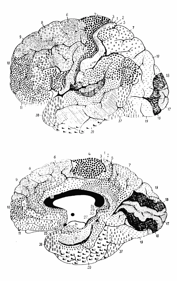
I will describe these areas in turn in relation to Brodmann’s description as well as the superficial landmarks. Figure 1 below shows many of the Brodmann’s Areas reproduced from Brodmann’s original 1909 work whilst Figure 2 shows some of Professor Cajal’s drawings of the structure of the cortex. Figure 3 shows the Brodmann Areas reproduced from a 1918 edition of Gray’s Anatomy whilst Figure 4 is a reproduction from a diagram in a paper by Hagmann and colleagues. In these diagrams, it is helpful to know that in most parts the Cingulate Cortex lies superior to the Corpus Callosum and indeed can be thought of as surrounding this structure (although the extremities of the Cingulate Cortex pass Anteriorly and Posteriorly underneath it).

Figure 1 – Cytoarchitectonics of human brain according to Brodmann (1909), Public Domain*, The Top Diagram is the Lateral Surface of the Cortex, The Bottom Diagram is the Medial Surface

Figure 2 – Three drawings by Santiago Ramon y Cajal, taken from the book “Comparative study of the sensory areas of the human cortex”, pages 314, 361, and 363, Public Domain*
Left: Nissl-stained visual cortex Middle: Nissl-stained motor cortex Right: Golgi-stained cortex

Figure 3 – The Anterior Cingulate Cortex, Brodmann Area 24, Derived from Gray’s Anatomy 20th Edition 1918 Lithograph Reproduction, Public Domain

Figure 4 – Hagmann et al, (2008), Extract from Figure 1 from Mapping the Structural Core of Human Cerebral Cortex, PLoS Biol 6(7): e159, Creative Commons 2.5 License
Turning first to Brodmann Area 23 (the Ventral Posterior Cingulate Area) the reader is directed to Figure 1. Broadly speaking, the area under discussion is contained within the Posterior Cingulate region which lies above the Corpus Callosum and would according to Brodmann’s description extend into the Isthmus of the Cingulate Cortex Corpus Callosum. This is because the Isthmus is shown in the diagram as directly in contact with the Precuneus along its inferior border. However the Precuneus is defined caudally by the Posterior-Occipital Sulcus which incidentally corresponds with the caudal limits of Brodmann Area 23 according to Brodmann himself. Rostrally Brodmann demarcates the borders as a transition with the Agranular intermediate Cingulate Area. In other words, Brodmann is defining the borders of BA23 by the microscopic agranular features of the cortex.
Now turning to Brodmann Area 31 (the Dorsal Posterior Cingulate Area) Brodmann describes this as occurring in the dorsal aspect of the Posterior Cingulate Cortex and arcing around Brodmann Area 23 until the Parieto-Occipital Sulcus. Brodmann’s description is slightly confusing in relation to the above diagram as he is familiar with the Isthmus of the Cingulate Cortex which he describes separately under the Retrospenial Region. Brodmann does describe a relation both to the Parietal Cortex and the Subparietal Sulcus but the description of these relations is rather vague.
Next looking at Brodmann Area 24 (the Ventral Anterior Cingulate Area) is essentially the anterior half of the Cingulate Gyrus located next to the Corpus Callosum with the exception of Brodmann Area 33. Caudally this extends to the transition zone defined by the change in granularity.
Brodmann Area 32 is the Dorsal Anterior Cingulate Area which extends to the Cingulate Sulcus Anteriorly as well as the Superior Rostral Sulcus ventrally.
Brodmann Area 33 is the Pregenual Area which is a thin strip of the Anterior Cingulate Cortex which lies adjacent to the Corpus Callosum through its course and passes deep into the Callosal Sulcus.
Brodmann Area 25 is the Subgenual Area. Brodmann’s microscopic analysis revealed a basic laminar pattern. The area extended from the Fornix to the Transverse Rostral Sulcus and inferiomedially is close to the Olfactory Trigone. This can be visualised relatively in Figure 3.
References
Brodmann’s Localisation in the Cerebral Cortex. 1909. Translated and Edited by Laurence J Garey. Springer. 2006.
*Public Domain in those countries where the Copyright term of the life of the author (Korbinian Brodmann 1868-1918) plus the additional country specific term has lapsed from Copyright at the time of writing
Appendix
Is it Time for Neuroscientists to Revisit the Brodmann Areas?
An index of the TAWOP site can be found here and here. The page contains links to all of the articles in the blog in chronological order. Twitter: You can follow ‘The Amazing World of Psychiatry’ Twitter by clicking on this link. Podcast: You can listen to this post on Odiogo by clicking on this link (there may be a small delay between publishing of the blog article and the availability of the podcast). It is available for a limited period. TAWOP Channel: You can follow the TAWOP Channel on YouTube by clicking on this link. Responses: If you have any comments, you can leave them below or alternatively e-mail justinmarley17@yahoo.co.uk. Disclaimer: The comments made here represent the opinions of the author and do not represent the profession or any body/organisation. The comments made here are not meant as a source of medical advice and those seeking medical advice are advised to consult with their own doctor. The author is not responsible for the contents of any external sites that are linked to in this blog.

[…] Smart Drugs & tDCS ReviewedShedding “Light” on the fMRI Debate with ofMRI.The Anatomy of the Cingulate Region: The Most Important 21 Pages In The Field of Neuroscience? Dr Ko… .recentcomments a{display:inline !important;padding:0 !important;margin:0 […]
LikeLike
[…] Corpus Callosum. I pointed out some interesting relationships with the structures in Figure 4 in a previous post which the reader is directed to when considering these […]
LikeLike
[…] The Anatomy of the Cingulate Region: The Most Important 21 Pages In The Field of Neuroscience? Dr Ko… […]
LikeLike
[…] The Anatomy of the Cingulate Region: Dr. Korbinian Brodmann – Part 9 […]
LikeLike