
Figure 1 –Dr Korbinian Brodmann, German Neurologist, Frontpiece of ‘Localisation in the Cerebral Cortex’, 1909, Public Domain*
Dr Korbinian Brodmann was a German Neurologist who undertook landmark research into the anatomy of the brains of many species including humans. The cornerstone of his work was a detailed microscopic study of the Cerebral Cortex which he related to the gross anatomical structures within the brain. His work has become extremely popular and stood the test of time with his Brodmann Areas remaining essentially unchanged since his 1909 landmark text ‘Brodmann’s Localisation in the Cerebral Cortex’. In the English translation, 21 pages are devoted to the study of the human Cerebral Cortex and these pages form the basis for the current localisation of the Brodmann Areas in humans. Brodmann’s coverage of other regions is covered elsewhere on this site (see Appendix). The last of the regions Brodmann described in humans is the Hippocampal Region. He described the following Brodmann Areas within the Hippocampal Region
Brodmann Area 27: The Presubicular Area.
Brodmann Area 28: The Entorhinal Area.
Brodmann Area 34: The Dorsal Entorhinal Area.
Brodmann Area 35: The Perirhinal Area.
Brodmann Area 48: The Retrosubicular Area.
These will now be covered in turn.

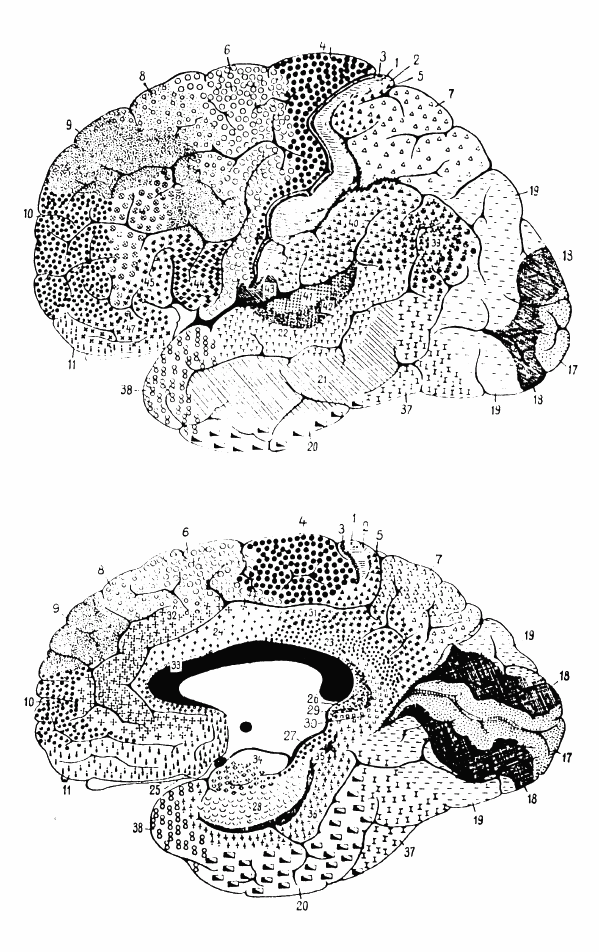
Figure 1 – Cytoarchitectonics of human brain according to Brodmann (1909), Public Domain*, The Top Diagram is the Lateral Surface of the Cortex, The Bottom Diagram is the Medial Surface

Figure 2 – Three drawings by Santiago Ramon y Cajal, taken from the book “Comparative study of the sensory areas of the human cortex”, pages 314, 361, and 363, Public Domain*
Left: Nissl-stained visual cortex Middle: Nissl-stained motor cortex Right: Golgi-stained cortex

Figure 3 – Stylised Diagram of the Hippocampus, Frank Gaillard, GNU Free Documentation License
Brodmann described the Hippocampal region as extending from the Hippocampal Sulcus (see Figure 3) to the Rhinal Sulcus.
Brodmann Area 27: The Presubicular Area. Brodmann describes the Presubicular Area as continuing directly on from the Subiculum (see Figure 3). He notes that there is a sharp demarcation in moving from the Subiculum to the Presubiculum (and references the change in cytoarchitecture seen in histological slides from the Kangaroo and Wallaby in a different section of the book). However he does not describe the nature of this demarcation in humans.
Brodmann Area 28: The Entorhinal Area. Brodmann describes this as lying adjacent to the Rhinal Sulcus. The anterior border is described as the ‘Temporal Incisura’. The term Incisura means notch and the Temporal Incisura is anatomically related to the Oculomotor Nerve. According to this surgical reference the Temporal Incisura is contained within a region described by the following
‘transversal line made in front of the cerebral peduncles and another line through the posterior border of the quadrigeminal plate into the anterior, middle and posterior parts‘
Brodmann suggests this is a vestigial remnant of the Posterior Rhinal Sulcus of other mammals. Brodmann describes the laminar pattern in the cytoarchitecture as characteristic throughout the species he examined. In the ventrolateral part he describes a change in the cytoarchitecture sufficient to enable him to describe the Dorsal Entorhinal Area of Brodmann Area 34.
Brodmann Area 34: The Dorsal Entorhinal Area. Brodmann describes this area simply as above and also as being medial to the Inferior Rhinencephalic Sulcus.
Brodmann Area 35: The Perirhinal Area. Brodmann describes this area as being limited to the Rhinal Sulcus. He also notes the absence of an inner granular layer and that this is the border between the Archipallium and Neopallium. Brodmann suggested that this area could be allocated to either although he seemed to favour the Archicortex in his 1909 work.
Caudal to the Perirhinal Area and lateral to the Presubiculum, Brodmann very briefly mentions the Retrosubicular Area (Brodmann Area 48).
Many of the Brodmann Areas can be seen in the schematic diagrams in Figures 4 and 5.

Figure 4 – Brodmann Areas 27, 28, 34, and 35 (visible on the medial view) Derived from Gray’s Anatomy 20th Edition 1918 Lithograph Reproduction, Public Domain

Figure 5 – Hagmann et al, (2008), Extract from Figure 1 from Mapping the Structural Core of Human Cerebral Cortex, PLoS Biol 6(7): e159, Creative Commons 2.5 License
References
Brodmann’s Localisation in the Cerebral Cortex. 1909. Translated and Edited by Laurence J Garey. Springer. 2006.
*Public Domain in those countries where the Copyright term of the life of the author (Korbinian Brodmann 1868-1918) plus the additional country specific term has lapsed from Copyright at the time of writing
Appendix
Is it Time for Neuroscientists to Revisit the Brodmann Areas?
An index of the TAWOP site can be found here and here. The page contains links to all of the articles in the blog in chronological order. Twitter: You can follow ‘The Amazing World of Psychiatry’ Twitter by clicking on this link. Podcast: You can listen to this post on Odiogo by clicking on this link (there may be a small delay between publishing of the blog article and the availability of the podcast). It is available for a limited period. TAWOP Channel: You can follow the TAWOP Channel on YouTube by clicking on this link. Responses: If you have any comments, you can leave them below or alternatively e-mail justinmarley17@yahoo.co.uk. Disclaimer: The comments made here represent the opinions of the author and do not represent the profession or any body/organisation. The comments made here are not meant as a source of medical advice and those seeking medical advice are advised to consult with their own doctor. The author is not responsible for the contents of any external sites that are linked to in this blog.

[…] blog???????????????? ?????????????????????????????????????????????????? ??????,????,The Anatomy of the Hippocampal Region: The Most Important 21 Pages In The Field of Neuroscience? Dr …?????? ?????????????????????????????????????????????????? […]
LikeLike
[…] The Anatomy of the Hippocampal Region: Dr. Korbinian Brodmann – Part 10 […]
LikeLike